This is one of the more complex cases I've worked on: the patient was missing bone across the entire span from the forehead down through the eye socket. From the CT scan, I segmented the defect in 3D Slicer, modelled the implant geometry in 3‑matic, and used Blender for additional sculpting and visualisation - building a plate that follows the patient's own skull curvature exactly, with a lattice structure for bone integration and fixation tabs positioned around the defect margin.
Defect mapping
Locating the defect in 3D
The affected region - highlighted here in pink - was isolated from the CT segmentation and checked from every angle before any implant geometry was drawn.
Implant design
From defect to plate geometry
The implant was designed to follow the defect margin precisely, with a perforated lattice body for tissue integration and fixation tabs placed around stable bone.
Final render - bone
Before and after, on the skull
The defect before design work began, and the same skull with the finished implant seated in place.
Before & after - soft tissue
What the implant restores
The forehead depression caused by the underlying bone loss, and the corrected contour once the implant is seated beneath the skin.
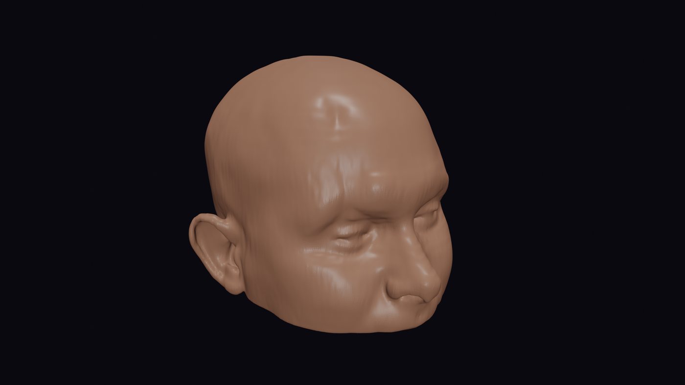
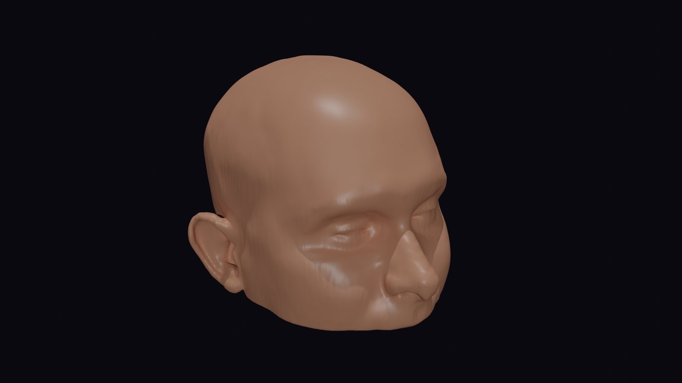
Every plate is drawn to the patient's own anatomy - the goal is never a generic fit, it's this exact skull, this exact defect.
Have a similar case?
Send over the patient's DICOM data and a brief description of the defect - I'll confirm feasibility within a few hours.
Send a message